Opuntia bigelovii structural Views

Anything relating to Cacti or CactiGuide.com that doesn't fit in another category should be posted under General.
User avatar
hegar
Posts: 4596
Joined: Mon Jan 23, 2006 4:04 am
Location: El Paso, Texas

Opuntia bigelovii structural Views

Post by hegar »

Two days ago I used my dissecting microscope, in order to figure out what kind of odd structures were present near the areoles of a Cylindropuntia bigelovii. I first thought, that I was looking at perhaps some kind of galls. However, cutting these off-white finger-like projections revealed to me, that they were solid, i.e. not caused by either insects or plant pathogens. They were also present near each areole examined. So I assume, that they may be extra floral nectaries. Is there anyone on this forum who could either tell me, that this is indeed the case or make another determination?
I am going to attach digital images showing these structures at 15x, 30x. and 40x magnification. Before I could take the digital images, I needed to remove some of the spines of the "cholla" cactus. So I went ahead and also photographed a piece of one of these spines, magnified 135x.
The plentiful barbs, which are arranged like fish scales are clearly visible. Those barbs are found with all members of the Cylindropuntia genus.
They explain, why the spine will easily prick your finger, but will be hard and very painful to be removed, ripping a larger hole than the diameter of the spine would suggest.

Harald

P.S.: I did label the images "O_bigelovii nectaries", because that made the most sense to me. However, I may be wrong with that assumption.
O_bigelovii nectaries x15.jpg
O_bigelovii nectaries x15.jpg (96.23 KiB) Viewed 3017 times
O_bigelovii nectaries x30.jpg
O_bigelovii nectaries x30.jpg (81.76 KiB) Viewed 3017 times
O_bigelovii nectaries x40.jpg
O_bigelovii nectaries x40.jpg (80.96 KiB) Viewed 3017 times
O_bigelovii spine x135a.jpg
O_bigelovii spine x135a.jpg (52.41 KiB) Viewed 3017 times
User avatar
ElieEstephane
Posts: 2909
Joined: Tue Dec 13, 2016 12:10 am
Location: Lebanon (zone 11a)

Re: Opuntia bigelovii structural Views

Post by ElieEstephane »

These are some scary looking spines
There are more cacti in heaven and earth, Horatio, than are dreamt of in your philosophy.
One of the few cactus lovers in Lebanon (zone 11a) :mrgreen:
User avatar
saboten
Posts: 178
Joined: Sun Nov 19, 2017 2:29 am
Location: Riyadh KSA

Re: Opuntia bigelovii structural Views

Post by saboten »

I don't have an answer about the nectary question, but thanks for uploading these pictures! I would love to see more magnified shots like this. Super fascinating =D>
User avatar
TimN
Posts: 3443
Joined: Mon Aug 06, 2007 9:01 pm
Location: Phoenix, Arizona USA

Re: Opuntia bigelovii structural Views

Post by TimN »

Nice pics! Never heard of extra-floral nectaries on any opuntioid, but I'll admit I'm not a diligent student of them. They sure seem to come close to the description.

Congrats for even getting that close to a piece of that plant.

Were the spines sheathed? They don't look like it in the picture.
Disclaimer: I'm in sunny Arizona, so any advice I give may not apply in your circumstances.

Tim
User avatar
ElieEstephane
Posts: 2909
Joined: Tue Dec 13, 2016 12:10 am
Location: Lebanon (zone 11a)

Re: Opuntia bigelovii structural Views

Post by ElieEstephane »

I just remembered, opuntia ficus indica has extra-floral nectaries. I often see the dripping and ants and bees come to feed on them. So i guess other opuntoids can have them too
There are more cacti in heaven and earth, Horatio, than are dreamt of in your philosophy.
One of the few cactus lovers in Lebanon (zone 11a) :mrgreen:
DaveW
Posts: 7399
Joined: Thu Dec 20, 2012 2:36 pm
Location: Nottingham, England/UK

Re: Opuntia bigelovii structural Views

Post by DaveW »

Many cacti have extra-floral nectaries, though we may not realise it.

There was an excellent article on them in the American Journal, reproduced in the link below. You have to scroll down a bit past all the headings before the article actually starts:-

https://www.researchgate.net/publicatio ... s_in_cacti
User avatar
hegar
Posts: 4596
Joined: Mon Jan 23, 2006 4:04 am
Location: El Paso, Texas

Re: Opuntia bigelovii structural Views

Post by hegar »

Thank you for all the interest you are showing in looking at the images above. I almost did not post them, but thought, that perhaps my guess would be validated. Thanks to DaveW it seems to have been correct. Because extrafloral nectaries exude a sugary solution, I had assumed, that the hyphomycete fungi were feeding on that substrate. They were not pathogenic, but the dark spores were quite noticeable. Once again, thank you DaveW for finding that article depicting these structures on cacti. I have recently joined ResearchGate and did not know, that this topic had been covered there. Of course, I downloaded the whole article and those beautiful images.

Well, Saboten, I shall post additional images of magnified cactus structures, if there is some interest. I had recently taken pictures of prickly pear stomates using a bit of clear nail polish, which I applied to the epidermis of the cladode. Then, after letting it dry, I peeled it off and placed it under my compound microscope. I tried to get open stomates, but each time I tried it, they were closed, despite having harvested the pad at night and kept in the dark. Perhaps my experiment did not work, because the plant was still dormant.

@ TimN: I did cut off the nasty spines with a pair of scissors. That allowed me to get close enough to take images through the lens of my dissecting microscope. The spines were sheathed. Image one does show a remnant of a sheath. With that microscope I can magnify objects 135x, which I did, when I wanted to show the spine surface with its barbs.

@ ElieEstephane: You are correct about the ferociousness of the spines. That cholla is supposed to have perhaps the nastiest barbed spines of any Cylindropuntia spp. I do have other chollas, including O. imbricata and O. leptocaulis. I can go ahead and photograph those spines too for comparison purposes. That might be an interesting study.


Harald
User avatar
hegar
Posts: 4596
Joined: Mon Jan 23, 2006 4:04 am
Location: El Paso, Texas

Re: Opuntia bigelovii structural Views

Post by hegar »

I am going to post digital images of the imprints made by stomates described in my prior text. As you can see, the stomates are closed. This could have been, because the Opuntia sp. plant was still in dormancy in February when I cut off the cladode, or perhaps because the photo was taken during the regular day time hours and the plant, having an internal clock closed the stomates, although I had kept the cladode in darkness from the time I cut it around 10 p.m. until I took the stomate imprints. I may run the same experiment again, because the prickly pear cacti are definitely actively growing and in the process of producing flowers now, just to see what the outcome is going to be this time around. I had hoped to see open stomates under my microscope cover slip. The images show the stomates at 200, 400, and 600 times magnification.

Harald
Stomata x200.jpg
Stomata x200.jpg (70 KiB) Viewed 2938 times
Opuntia stomates x400.jpg
Opuntia stomates x400.jpg (91.29 KiB) Viewed 2938 times
Opuntia Stoma x600.jpg
Opuntia Stoma x600.jpg (74.07 KiB) Viewed 2938 times
User avatar
ElieEstephane
Posts: 2909
Joined: Tue Dec 13, 2016 12:10 am
Location: Lebanon (zone 11a)

Re: Opuntia bigelovii structural Views

Post by ElieEstephane »

hegar wrote: Sat Apr 06, 2019 2:01 am I shall post additional images of magnified cactus structures, if there is some interest.
There definitely is!
There are more cacti in heaven and earth, Horatio, than are dreamt of in your philosophy.
One of the few cactus lovers in Lebanon (zone 11a) :mrgreen:
User avatar
autodikaktos
Posts: 57
Joined: Sun Dec 30, 2018 7:06 pm
Location: Arizona, USA (Zone 7B)

Re: Opuntia bigelovii structural Views

Post by autodikaktos »

Thank you so much for creating this thread and posting these photos. I very much hope to see more. I mean, *really* fascinating stuff. I'd been hoping for a follow up from the previous bigelovii thread. Ever since I read it I have been nagged by these non-verifiable memories of seeing those little nubs in Cylindropuntias in habitat. After hitting up some of the academic stuff, go figure, Cylindropuntia acanthocarpa and imbricata both have them, and ants are all over them. And as I'm writing this I'm remembering that I used to be curious on the crystallized "stuff" on the areoles of new pads on different Opuntia species, which also tended to attract a lot of ants. I wish the ants that spend all their time moving aphids around on my plants would bother and do a little defense as their mutualist wild counterparts do. :roll:

Although I only read it in an abstract, it would seem that at least one of the southwestern Opuntias have ephemeral nectaries whereas the Cylindropuntias maintain theirs. Seriously, moments like these are why I love this hobby so much. Really appreciate sharing your efforts with us.
"yep, son, we have met the enemy and he is us." ](*,)
User avatar
Licespray
Posts: 352
Joined: Mon Mar 19, 2018 10:26 pm

Re: Opuntia bigelovii structural Views

Post by Licespray »

ElieEstephane wrote: Fri Apr 05, 2019 2:41 pm I just remembered, opuntia ficus indica has extra-floral nectaries. I often see the dripping and ants and bees come to feed on them. So i guess other opuntoids can have them too
I’ve observed ants feeding on what I believe are extra-floral nectaries on Cylindropuntia imbricata here too.

Searched and apparently I did take photos.
Attachments
D39D292B-A515-433C-AE0B-4F2F34B68E81.jpeg
D39D292B-A515-433C-AE0B-4F2F34B68E81.jpeg (102.48 KiB) Viewed 2861 times
Ferocactus best cactus :mrgreen:
User avatar
hegar
Posts: 4596
Joined: Mon Jan 23, 2006 4:04 am
Location: El Paso, Texas

Re: Opuntia bigelovii structural Views

Post by hegar »

Hello Elie, autodikaktos, and Licespray,
thank you for the encouraging comments. Not only do you get to see things, which you might not have any chance to do otherwise, but I too profit and learn new things.
Now, I am willing to use the skills and equipment I do have - in a limited way, however - to find out things that are of interest to the cactus-loving community here on the CactiGuide blog. It would help though, if someone were to give me a few suggestions.
I had already mentioned previously, that I could repeat the stomate experiment, in order to find out, if the cactus closes the stomates when the plant receives light or if it does it, even if it is kept in the dark, but in reality it is daytime. That would indicate, that an internal clock tells the plant to shut the stomates.
I am also already in the process of photographing quite a number of cactus seeds. My goal is to show the whole seed, its hilum (seed scar) and the testa (seed coat) under various magnification levels. I do intend to have this finished by the time I retire, which may be in about one year. After that I will no longer have the use of the microscopes, unless of course someone gives me a special authorization based on my long tenure.
Once I am done with that project, I would gladly make this image material available to everyone who is interested in it. Because of the number of images and the sheer size of the document I could not post this work here, but it could be placed into a drop box.

Harald
User avatar
autodikaktos
Posts: 57
Joined: Sun Dec 30, 2018 7:06 pm
Location: Arizona, USA (Zone 7B)

Re: Opuntia bigelovii structural Views

Post by autodikaktos »

Again, for I believe the third time in two days, thank you so much for your efforts and your participation in the "hobby," although that word does not do justice to the subject. So many of us just don't have direct access to the academic articles and materials, so its just an absolute treat to come here and see this kind of thing at the amateur level. At least personally I would have interest in everything you have mentioned. Seeds, spines, and stomata would all be great things to see and understand more. My apologies that I don't have any suggestions that would prove or disprove a hypothesis, but I am curious at seeing such things as the makeup of succulent root systems, the bases of glochids... Also cactus leaves... particularly those of opuntoids (both cotyledons and the ephemeral leaves on the new cladodes).

I do wish you luck for the seed project in particular, and hope to be able to peruse the work at some point in the future.
"yep, son, we have met the enemy and he is us." ](*,)
DaveW
Posts: 7399
Joined: Thu Dec 20, 2012 2:36 pm
Location: Nottingham, England/UK

Re: Opuntia bigelovii structural Views

Post by DaveW »

Joel Lode has done quite a bit of Cactus seed photography as has Dr Detlev Metzling. Problem can be it often comes somewhere between low magnification microscopy and higher magnification macro photography. Also these days of digital photography, either using a microscope or with macrophotography you need to focus stack to get all seed in focus. In the old days with film you were restricted to merely balancing reasonable depth of field against diffraction and putting up with a softer image to get more in focus, since focus stacking only came about with digital photography and computers.

Scroll down this link.

http://cactus-aventures.com/Taxonomy/Td ... ctENG.html

Some of Detlev Metzings here:-

http://forum.bcss.org.uk/viewtopic.php? ... 0&start=10

I am in the preliminary stages of setting up to do some pictures of seed for a friend using a macro setup. He is using a microscope too but says the magnification is too much for what he wants as he often wants more than one seed in shot, but a small group with one standing on end to show the hilum.. This was just a quick test to see if I could manage to set the pin head sized Thelocephala seeds to stand up on double sided Sellotape. But as I have had flu at the moment so not being able to see tiny seed with bleary eyes it will have to wait a week or so until I am back to normal.

The idea is the setup in the picture below with a milk white diffuser made from a plastic bottle neck lit by two flashguns and then do around a 12 image focus stack using the StackShot rail at the back that can automatically move the camera in sub millimeter increments if needed. Camera goes on right top but had to use it to take the picture.

seed-setup.jpg
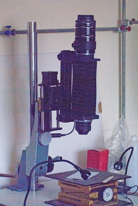
seed-setup.jpg (119.68 KiB) Viewed 2817 times
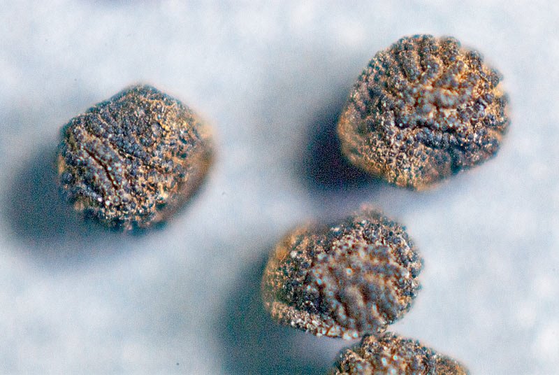
These were just preliminaries used to see if I could get the magnification required and obviously focused a little too far in to start. Different background colours may be used but of course any background colour can be changed in post processing anyway. I am just proceeding one step at a time to refine the setup. Of course at these magnifications you cannot stop down far as in normal macro photography since diffraction rears it's ugly head, softening the image and the lens had to be used around f8-f5.6 and the image focus stacked. Must be a doddle photographing something as large as Opuntia seed compared to Thelocephala's, and Aztekium's would be even worse but then a microscope would be better, but you would still probably need to focus stack to get all of the seed in focus at once in the picture.

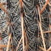
These have not been focus stacked since that would have had all the seed in focus, they are just a single trial shot which shows how small the depth of field is with a seed only a couple of millimetres in diameter. At those magnifications the depth of field can often only be as deep as the thickness of a piece of writing paper.

seedRMF2.jpg
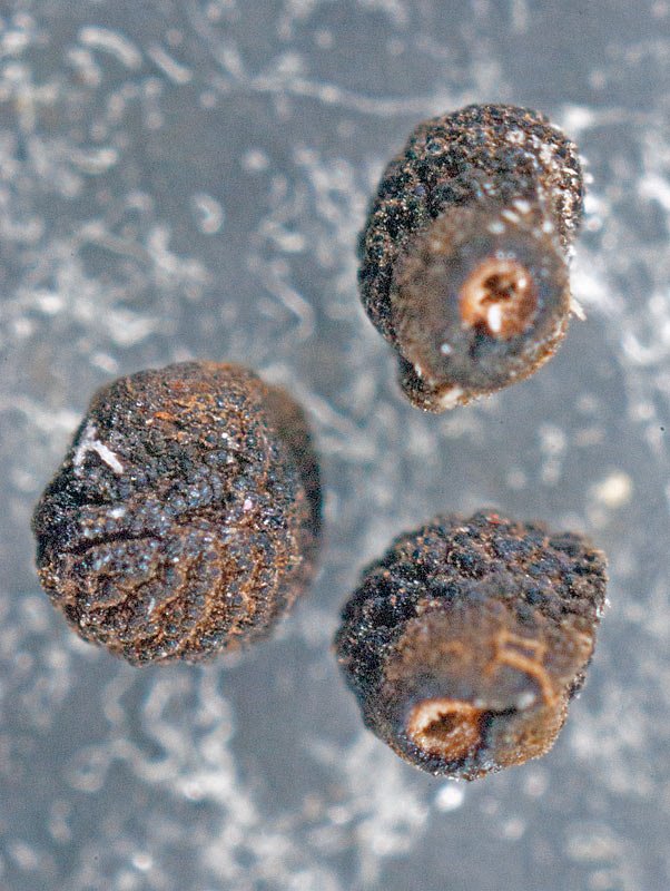
seedRMF2.jpg (114.76 KiB) Viewed 2817 times

seedx.jpg
seedx.jpg (96.46 KiB) Viewed 2817 times
Any ideas how to clean the brown material off such tiny seeds.

For those who do not know what focus stacking is and why often at higher magnifications enlarger lenses as on my setup can be better than a normal macro lens see:-

http://extreme-macro.co.uk/focus-stacking/

Now you know why many try it and give up! :lol:
User avatar
hegar
Posts: 4596
Joined: Mon Jan 23, 2006 4:04 am
Location: El Paso, Texas

Re: Opuntia bigelovii structural Views

Post by hegar »

Hello autodikaktos, hi DaveW,
thank you very much for your input. I am by no means a true scientist and oftentimes tell people, whenever I do give a PowerPoint presentation, that I am as good a plant pathologist as the wizard was one in the "Wizard of Oz" movie. So you cannot expect any breakthrough new knowledge from the world of science from me. I am just fortunate, that I do have pretty decent equipment that I can put to use, which allows me to see things that most people would not be able to spot, i.e. two pretty good microscopes - with the dissecting microscope being capable of 135x magnification - and also a camera system and software, which does allow photo stacking (Helicon Focus).
As far as cactus seed photography is concerned, I do have a nice book at work, which does show the same things I want to photograph before retiring. However, that work I cannot match or even get close, because the images were taken with a scanning electron microscope at magnifications I can only dream about. There was also an article I read about using low power compound microscope photography to photograph seeds and getting a better image than when using my high-powered dissecting microscope. That, however, requires some tinkering with the equipment and I am the last person on this planet who should be trusted with that task. So I have to make do with what I have right now. All I intend to do is to create some plates, which show the whole seed near a ruler with mm increments, the hilum highly magnified, and the testa at 100 or 135x magnification. I am not going to provide any text, only the name of the plant from which the seed was obtained. The plants I shall cover are pretty much all from the Chihuahuan Desert. So there will be neither South American cacti seed, nor seed of rare North American cacti presented. The latter, because I do not have seeds of them. I do have most of the cactus species seed from the Big Bend area, which were supplied to me by a professor, who works at Sul Ross State University.
Dave, I shall of course read everything that you have supplied me with on this blog, in order to see what I can apply to the task at hand. You are truly a fountain of information and I am grateful for the time and effort you extend to help anyone out who is looking for assistance. So I do hope, that I become a little more enlightened and educated by what you did provide this time around. Thank you once again!

Harald
Post Reply